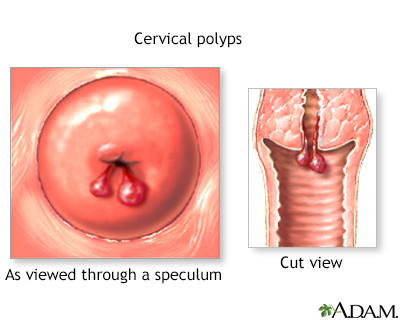
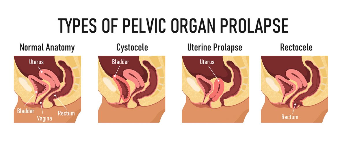
A. Cystic content was haematic. B: Hydatid membranes have the color red


Cystic liver lesions: a pictorial review, Insights into Imaging

Cardiac MRI images. A, Steady-state free precession sequence showing a
Hydatid Disease: A Pictorial Review of Uncommon Locations
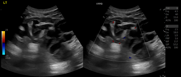
Hydatid disease, Radiology Reference Article
Manual of diagnostic ultrasound - World Health Organization

Cardiac MRI images. A, Steady-state free precession sequence showing a
Hydatid Disease: A Pictorial Review of Uncommon Locations

The Orbit, Including the Lacrimal Gland and Lacrimal Drainage System

Case Report: Infected primary hydatid cyst of the

PDF) Imaging characteristics of three primary muscular hydatid cyst cases with various patterns

